Hardware and software of the brain
The main approach to creativity remains ancient trials-and-errors method. For computers, more orderly variants such as full scan of possible solutions are used. In any case, the processor should prompt some actions, then assess their results. That's exactly what the limbic system does. Looks like the amygdala is a block of system-level memory similar to the basal ganglia. Only they keep elementary programs for sensorimotor coordination while the amygdala keeps genetically predetermined states of the brain itself such as fear or aggression. Human emotions correspond to processor instructions of computers, only the brain doesn't use the Von Neumann architecture. It uses a finite-state machine which has elementary states rather than actions. This approach may be very powerful. It is well known that the brain parts tend to have connections according to the principle of all-to-all. Not all of them are used at once. Instead, only a fraction is employed when necessary. This means that you can dynamically create different working machines for different circumstances.
On the other hand, the hippocampus has all the necessary means to assess the results. It receives input from major sensory channels and can generate sharp pulses of activation at the output. Again, this works differently. When you write a control program, you would create a variable to measure the assessment. Say, in the range [-1,1]. The program will input data, process it, set that variable, then use it for decision making. Looks like the brain has not such a separate variable. Instead, associative memory immediately links an input situation to the appropriate emotional reaction such as attraction or aversion. That's why negative emotions are harmful. You get tensed and if this tension has no exit, you must contain it. That leads to double tension and quick tiredness.
Other parts of functional architecture
If the neocortex is the associative memory of the brain and the limbic system – its processor, the next step would be to describe Input/Output system. In contrast to PC, human memory is anatomically partitioned and different parts are assigned for different purposes. Comparison of human and computer I/O systems may be as lengthy as the preceding comparison of central processing, but now we will just mention that they are very different as well and concentrate on the first. In particular, I/O system of PC was very asymmetric. These computers had quickly got a graphical display with a complicated controller and a video card, but the main input remained a keyboard and a mouse. When decent video input became available, PC itself already lost its market share. Output subsystem was obviously better developed here and the term "programming" reflects this. It refers more to generating output actions and pays less attention to analysis of input data.
In humans, the main input comes from vision and the main output goes to muscles via the motor system. Both are approximately equally developed. They have cortical and subcortical representation of comparable complexity. The human sensory system has several different receptors (vision, hearing, etc.) with separate sensory channels. At the top, all of them converge in the TPO (Temporalis-Parietalis-Occipitalis) region of the neocortex which resides between cortical representations of different sensory modalities. The TPO zone obviously syntheses an abstract picture of the world and is also related to human consciousness.
While the sensory neocortex occupies the back half of the brain surface, the front half is designated for motor output and actions in more general sense. Humans have only one channel of external output – muscles, but the overall output system is slightly more complicated than its input counterpart. It includes the cerebellum sometimes called a small brain inside the big one. The cerebellum may be considered as a close analogy of a video card in PC. It has its own cortex and subcortical nuclei that is seemingly memory and a processor, but is used for motor tasks.
At last, yet another part of architecture. The reticular formation has a few distinct features. It is located among the most ancient parts of the brain, is large, but has virtually no internal structure, and has diffuse output to other parts. This is an activating block of the brain. We could outline the analogy with the power unit of PC, but here the difference is probably the largest. The nervous system has no separate power supply at all. Instead, each neuron generates energy for itself. Nevertheless the On/Off switch is necessary. It comes in the form of the sleep/awakeness cycle. In contrast to computer electronics, neurons are heavily dependent on biochemistry which produces a lot of waste and requires regeneration of stock substances. That's why we sleep every day and the reticular formation controls this cycle. Moreover, it can fine-tune its output so as not just to turn the whole brain on or off, but to do it separately for different structures. The reticular formation not only sends output, but also receives input from other parts. This supports the principle of a dynamic processor discussed previously in connection with the amygdala.
That's all about the core functional architecture of the brain. Now let's formalize it in a concise description.
Formal neurocomputer
This machine is created from blocks of associative memory linked in certain order. Each block has input and output in the format of 2D or 3D image plus a control signal that switches the mode of operation – read out, write in, or inactivate. In the first mode, the block uses the input image as a key to retrieve an output one. In the second – memory remembers the association between 2 images.
The machine is connected to the external world by input and output. Both have hierarchical structure with several tiers and maybe several parallel channels which converge from periphery to the centre. The main difference is the direction of information flow. The sensory system gathers data from various receptors and creates an integral picture of the world in the central representation. The motor system gets a general idea of the action in its central representation, then implements it in concrete actions of various effectors.
Operation of the machine is determined by interaction between central images of sensory and motor systems. A sensory image serves as a key to the motor block. Associative memory generates an idea and while this idea is being realized, the associative pathways between the upper levels are inactivated. As soon as the action is completed, they are activated again, but now the content of sensory operative memory is already changed. Two sources are possible. The action may change something in the environment or data may be written directly into the sensory memory in the course of internal exchange. Hence the key will be different and associative memory will generate the next insight. The cycle repeats. So the work of this machine is based on chain reactions.
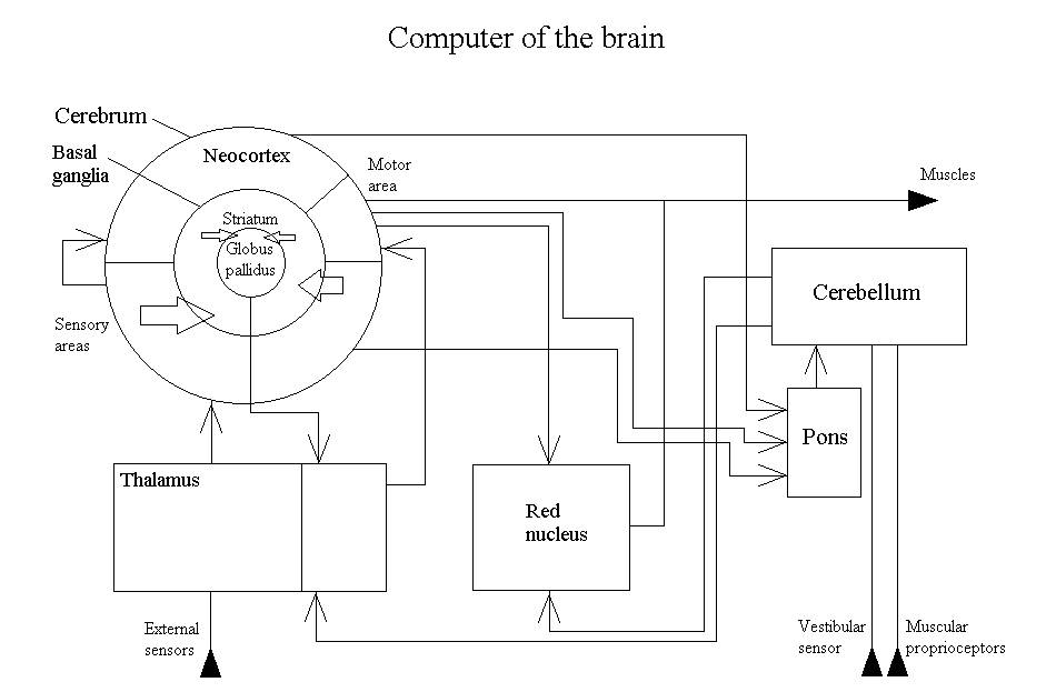
Fig. 7.
This scheme is compiled from the most reliably described links between various brain regions. How to understand its operation? The main problem here is multiple feedbacks. The common method in such cases is to break the loop. Then, operation of the open circuit will become obvious. So we need to remove some links from this scheme, but which specifically? The red nucleus is a clearly supplementary structure because the motor cortex already has direct output to motoneurons. So we will throw it out together with the cerebellum. Functions of the latter are well established. It provides muscular coordination and fine-tuning. If it is damaged, patients still cam move, but their movements become more primitive, rough, and non-coordinated. The cerebellum is a typical "improvement".
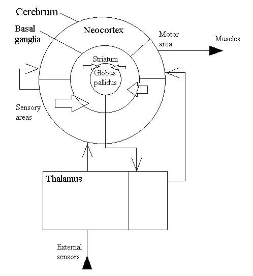
Fig. 8.
This picture still may be further simplified.

Fig. 9.
The upper link on the picture is a well known reflex. The link through the basal ganglia is a bit more complicated. It combines the previous principle of stimulus-reaction with sensorimotor coordination. The last detail comes from the fact that muscular actions change the environment and these changes will be perceived by external sensors.
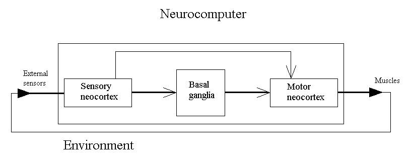
Fig. 10.
This device is not so weird as it may seem at the first glance. For illustration, let's demonstrate how to implement common linear programming here. Suppose you have some algorithm, that is a definite sequence of actions. Just associate each one with the corresponding number in the sequence and remember these associations. By the way, memorizing may be performed in any order. Next, you will need a small system program. Namely, you should have a sensory image which keeps the number of the current operation. That is you emulate the register of a common processor. Increment it after each action. Now use this image as a key for sensorimotor associations and you will be able to run common programs on this computer.
Processor of the brain
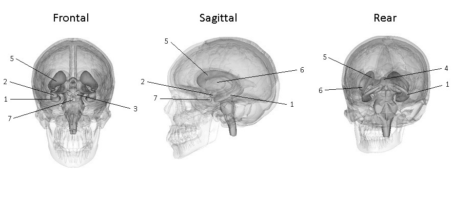
Fig. 11.
Limbic system.
1 – Hippocampus. 2 – Amygdala. 3 – Mammillary body. 4 – Fornix.
Basal ganglia.
5 – Caudate nucleus. 6 – Lentiform nucleus.
Other.
7 – Pituitary gland.
Like a personal computer, the human brain also has memory and a processor, although they operate and interact quite differently. Neurocomputing does not use linear memory containing bytes or machine words which may be accessed one by one. Instead, it relies upon associative memory that keeps bytes in synapses, but they can't be immediately read out. Humans use a different principle. Memory bytes form a distributed hologram which is only an instrument to retrieve data. The output comes in the form of an image encoded by a current spike rate distributed over some neural net. Again, in contrast to a computer, each neuron outputs not binary data of 1 or 0, but a real value in some range, say [0, 1]. This principle is used both in memory and a processor; they are different only by the architecture. Human memory is located in the neocortex which is a large 2D structure packed into the skull. It has 6 layers of neurons of different types which are basically the same throughout the whole of its area, but this area is genetically partitioned according to the functional principle. The frontal half of the neocortex works mainly for output, the rear – for input. Many different cortical fields are interconnected by direct axonal projections. This system alone already can work like computer software. When some portion of data comes to a sensory input, this is 1:1 as an event in a computer. This event produces activation which spreads to a different field, undergoes various processing, and finally may generate a muscular action. The problem is that this flow is not synchronized, no single clock generator. Also, brain connections tend to be organized by the principle of all-to-all or one-to-many. The uncontrollable spread of activity will lead to informational overflow. Obviously, for a given event, only a fraction of all fields and their links should be used. All the rest need to be inactivated. This is the main function of the brain processor – the limbic system.
In a computer, the processor has a definite set of instructions and handles data loaded from memory. In the brain, data are processed immediately in memory. The processor only starts and stops this process. Nevertheless, it has some instruction set too. The limbic system generates emotions. Motions are activation of muscles, emotions – similar activation of internal brain structures. Like the neocortex, parts of the limbic system also keep various images, but these images encode patterns of activity for other parts of the brain. Some of them need to be boosted, others – suppressed. Such emotional patterns may be learned, but also may be inborn basic distributions of strong reactions.
Another important function of the limbic system is related to operative memory. Associative memory does not keep the image itself – only the ability to evoke it. Nevertheless, neural nets can retain images too. For this purpose, they build a sort of a trigger system using positive biofeedback. In this case, the output of some internal block is supplied to its input starting recirculation in the closed loop. As soon as some pattern gets into this system, it becomes locked and can exist for a prolonged time even if the source was turned off. The limbic system was initially called the Papez circuit. It and the adjacent basal ganglia have many such loops which may be employed in various cases. Usually, some cortical field sends output to the limbic system and also receives input from it. Obviously, when some image is reverberating through the limbic structures, they can control it. That's how this system manages operative memory.
A well known function of emotions is assessment. In fact, most of them are broadly divided into 2 categories – positive and negative. The strength of emotion provides a value for this sign. Suppose you need to write a program for monitoring of some object. How would you do that? Probably you will define an appropriate variable, write the evaluation there, and use it for decision making when necessary. Seemingly the limbic system operates differently. The assessment is not kept separately but appears immediately at the output as the aforementioned control signal. That is, if the results of some actions are negative, the aversive emotion will immediately suppress that activity.
2 key blocks of the limbic system are the amygdala and the hippocampus. They are very different and seemingly complement each other. The amygdala is a typical subcortical structure. It is linked to generation of very basic states of the organism as a whole – such as fear and aggression. The hippocampus is the archicortex. This is a small piece of cortical matter which emerged early in evolution and has just 3 layers. Thus it should perform some important functions. An interesting theory of the hippocampus is as follows. What you usually see in illustrations (CA1, CA3, etc.) is a cross-section. The hippocampus as a whole is a tube or more precisely – a long cone in correspondence to its name Cornu Ammonis. Functionally, the hippocampus generates a 1D image that is a vector. The index of this vector goes along the tube while its cross-section provides a pathway for data rotation. Input comes from the entorhinal cortex, output returns back to it. That is, the entorhinal cortex may be a memory buffer which can receive an image from a wide variety of cortical regions, then keep it for temporary dynamical storage via reverberation through the hippocampus. Thus, the hippocampus may work as famous operative memory. Note that it can't store data because it is 1D, while cortical images are usually 2D. Only plays some important role in this storage. In addition to that loop, another major output of the hippocampus goes via the fornix to several small (0D that is scalar) areas of the brain. Through the mammillary bodies, the hippocampus can control the thalamus. It also sends output to the septum (a pleasure zone). There are several such spots in the brain (the substantia nigra, ventral tegmental area). They use the neuromediator dopamine which can control formation of long-term memory.
Конец ознакомительного фрагмента.
Текст предоставлен ООО «Литрес».
Прочитайте эту книгу целиком, купив полную легальную версию на Литрес.
Безопасно оплатить книгу можно банковской картой Visa, MasterCard, Maestro, со счета мобильного телефона, с платежного терминала, в салоне МТС или Связной, через PayPal, WebMoney, Яндекс.Деньги, QIWI Кошелек, бонусными картами или другим удобным Вам способом.
Вы ознакомились с фрагментом книги.
Для бесплатного чтения открыта только часть текста.
Приобретайте полный текст книги у нашего партнера:
Всего 10 форматов

